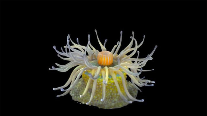
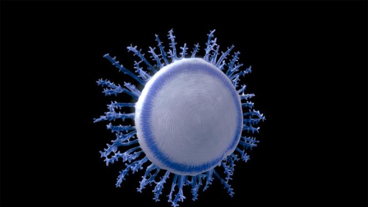
When James Hanken became the director of Harvard’s Museum of Comparative Zoology (MCZ) two decades ago, the museum’s hundreds of glass models of sea invertebrates, purchased in the late nineteenth century from the father-son pair of glass artists Leopold and Rudolf Blaschka, were hidden away in storage and in poor condition. The models—such as a translucent, tentacled sea anemone typically found in the Western South Atlantic, 10 centimeters wide and almost glowing pale yellow and green; or a sparkling seven-centimeter-diameter, Porpita porpita, a collection of hydroids that has the appearance of a light-blue button with darker, branch-like tentacles extending from its periphery—were gathering dust, and some were broken. The MCZ embarked on a conservation project, and now many of the glass sea invertebrates are on rotating display, though physical access is highly restricted. But in recent years, the entire world has become able to see and virtually “handle” some of the Blaschka models by spinning the life-like glass on a computer screen, thanks to new 3-D modeling techniques described in a recent study co-authored by Hanken.
Leopold Blaschka, on a trip to the Americas in 1853, became fascinated by the marine life he saw: “[T]here emerges all around in various places a flashlike bundle of light beams, as if it is surrounded by thousands of sparks, that form true bundles of fire and of other bright lighting spots, and the seemingly mirrored stars.” He found these bioluminescent, translucent creatures were glass-like in their beauty and thus sought to capture them in glass models, working meticulously from drawings and wet samples. His son joined the business in 1876—and business was prosperous, as the Blaschka models filled a need among nineteenth-century museums for sea invertebrates, because live specimens were difficult to preserve. Some of the techniques the Blaschkas used to accurately model marine life in glass remain a mystery to this day. And though many of the more than 10,000 models of sea creatures the father-son pair produced have been lost, Harvard maintains the second largest collection (after Cornell) in the world.
It was the Blaschkas’ story that drew Peter Fried, the study’s lead author and a professor of applied physics at NYU, to the sea invertebrate glass models five years ago. “I was taken by [the models’] beauty and craftsmanship and the whole story behind them,” he says. “On a lark,” Fried reached out to Cornell, expressing interest in rendering the glass models, and Cornell agreed. Soon after, he attended a conference on 3-D scanning. Several companies and experts told him scanning the Blaschka models would be almost impossible—which only heightened his interest.
Traditional 3-D photography, called photogrammetry, involves photographing an object from various angles, identifying features that look the same across photos, and using those constants to reconstruct a 3-D model from all of the images. The Blaschka models, however, are glass—their reflections and glare change based on the position of the observer and the light, which means photogrammetry struggles to identify the necessary constant features. For some of the more difficult models, such as the sea anemone, however, the researchers were able to optimize lighting to minimize or maximize specular reflection, allowing them to create a full 3-D rendition through photogrammetry alone.
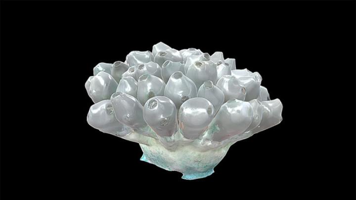
But other models could not be captured with photogrammetry, due to a combination of hidden surfaces, transparent sections, and significant reflections. The football sea squirt, for instance—which resembles a bouquet of silver-grey olives on toothpicks—posed such difficulties. But the researchers knew of another imaging technique, x-ray computed tomography (CT), which can model reflective or transparent objects. A CT scan, which the MCZ uses often to scan museum specimens, takes a series of cross-sections of a specimen and compiles them. X-rays have trouble detecting very thin surfaces or coatings, though, meaning CT scanning couldn’t pick up the Blaschka models’ almost ethereal colors, but combined, CT scans and photogrammetry are complementary.
The paper shows several digital renditions of the football sea squirt. One, taken with x-rays, captures the bulbous shape but not the white-green paint on the model’s underside. Another, made with photogrammetry techniques, has a distorted shape. When integrated, the photogrammetry and x-ray-generated images created a near-perfect model.
Fried has created 3-D models of more than a dozen of the Blaschka models, viewable on the website Sketchfab. The combination of photogrammetry and CT scanning has made the Blaschka models accessible to researchers around the world—and, Hanken adds, may be useful to museums with models that are similarly too fragile to put on display. In digital form, however, visitors can for the first time interact with these unique objects, as though turning them over in their hands.
